Anemia in pigs caused by Mycoplasma suis
Causes
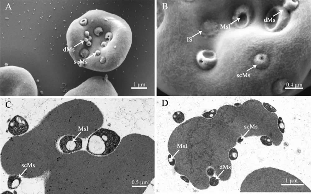
The disease is caused by the bacterium Mycoplasma suis, belonging to the genus Mycoplasma. It is spherical or oval in shape, with a size of approximately 0.8 μm – 2.5 μm. Because Mycoplasma suis parasitizes on and within red blood cells, infected pigs exhibit a decrease in the percentage and number of red blood cells, hemoglobin, glucose, and iron concentrations, while bilirubin levels in the blood increase, leading to acute or chronic anemia in infected pigs, reducing productivity, increasing the rate of secondary infections, and causing death due to superinfection in all groups of pigs infected with M. suis (Ritzmann et al., 2009).
Epidemiology of the disease
The disease occurs in pigs of all ages. Studies worldwide show that infection rates vary depending on geographical region and age of the pigs, with particularly high rates of infection in sows on farms. In Germany, up to 40.8% of farms were infected with M. suis, with approximately 13.9% of young pigs (20-30 kg) testing positive for M. suis (CSIRO, 2012). According to Stadler et al. (2019), the rate of newborn piglets testing positive for M. suis before suckling was 14.35%, while 31.25% of sows tested positive. In contrast, the rate of M. suis-positive pigs in Australia is very low, ranging from 4.29-6.45% (CSIRO, 2012). In France, over 50% of sows tested positive for M. suis across all litters.
The disease is primarily transmitted through blood, not through semen or saliva. The early stages of piglet life – when technical procedures such as clipping teeth, tail docking, castration, and vaccination are performed – pose a significant risk of disease transmission between herds if proper hygiene and disinfection are not maintained.
According to research by Zhongyang et al. (2017), the infection rate of M. suis fluctuates with the weather, increasing during transitional seasons and the dry season. This is also the time that creates favorable conditions for blood-sucking insects such as flies and mosquitoes to multiply.
External stress factors such as weather, environment, and animal health status are risk factors that exacerbate M. suis-induced disease. Infection from outside mainly occurs through the introduction of infected pigs into the farm.
Symptoms
The disease usually appears in two forms: acute and chronic.
* Acute form:
+ For suckling piglets
Pigs are stunted, pale, whitish, and emaciated.
They have low uniformity, poor growth, and vary in size.
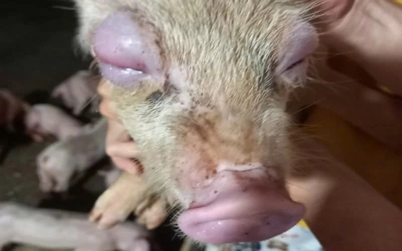
The ear rims may be purplish-blue, and the eyelids may be swollen.
Weakness in the legs may occur, piglets may tremble, walk unsteadily, and have convulsions due to hypoglycemia.
+ For weaned piglets:
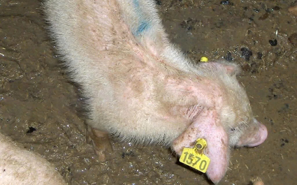
Fatigue, lethargy, reduced appetite, slow growth.
Pale skin and mucous membranes, poor physical condition, stunted growth, rough coat.
Rapid breathing, abdominal breathing.
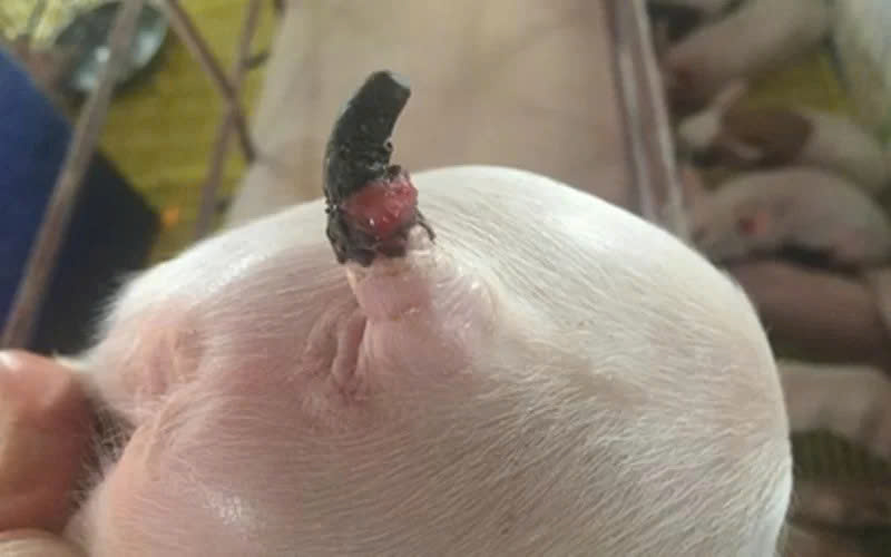
Necrosis of the ear and tail tips.
Pigs with tail tip necrosis.
Pigs stunted, slow-growing, rough coat.
+ For sows:
High fever (40-42°C), increased respiratory rate, decreased appetite
Anemia, pale skin (jaundice), loss of milk
Delayed estrus, miscarriage or premature birth
Reduced mating success rate, reproductive dysfunction
Pigs born thin and stunted
* Chronic Form
Antibiotic treatment is insufficient to completely eradicate M. suis from infected pigs, leading to chronic infection. Pigs exhibit poor health, jaundice, stunted growth, dry skin, rough coat, and immunosuppression, making them susceptible to respiratory and digestive diseases, thus increasing treatment costs.
Pathological Lesions
Pale skin and mucous membranes due to the breakdown of red blood cells, accumulation of by-products in the liver, and the production of bilirubin.
The blood is thin, red blood cells are deformed into star shapes, and their biological function is reduced or lost.
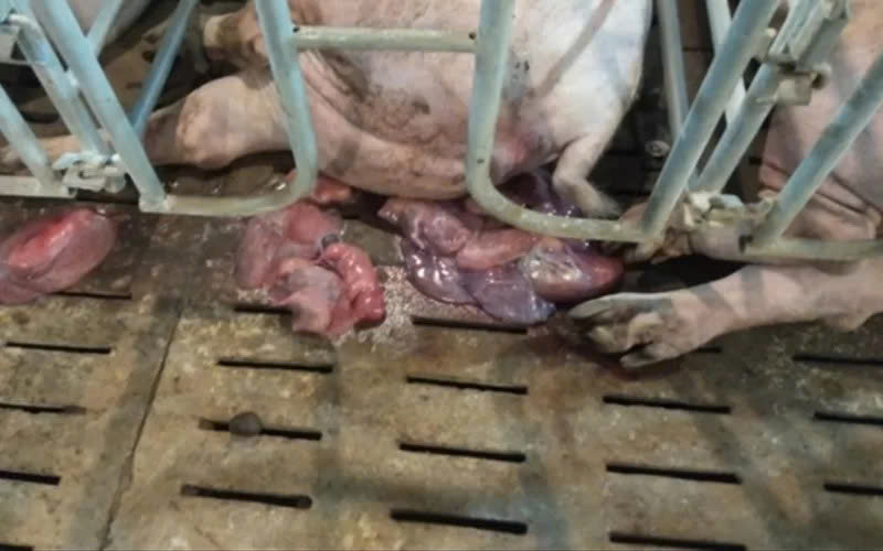
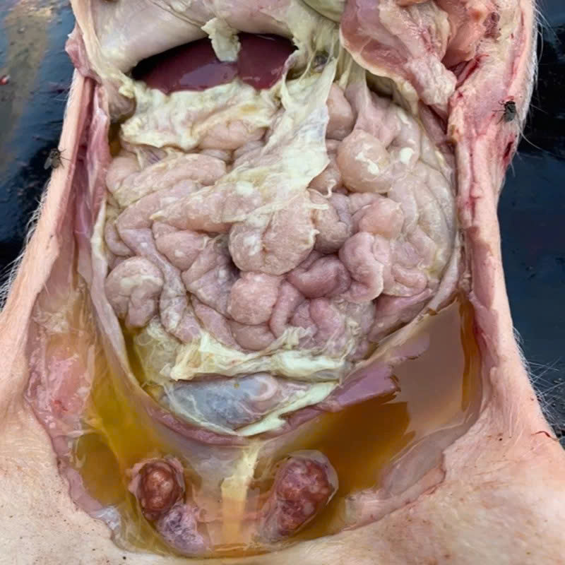
In weaned and young pigs, skin ulcers may appear on the edges of the ears, tail, and snout. Post-mortem examination may reveal lesions such as pericarditis and effusion in the abdominal cavity, chest, pericardium, thin heart walls, flabby heart, swollen spleen, and swollen lymph nodes.

Diagnosis
Clinical Diagnosis
Based on the trembling symptoms of newborn piglets. Additionally, it is based on signs such as pale and white skin, dry, ruffled hair, swollen eyelids, and ulcers on the edges of the ears and tail in suckling pigs, post-weaning pigs, and young pigs.
Non-Clinical Diagnosis
Diagnosis is primarily based on testing for the presence of M. suis in the blood. To test for M. suis, a blood sample with anticoagulants is taken for Giemsa staining to observe M. suis on red blood cells, or a PCR reaction is performed to identify M. suis in the blood sample.
Differential Diagnosis
Differential diagnosis should be made with other diseases that also cause jaundice and skin necrosis, such as Leptospira infection, PCV2-associated disease (PCVAD), Glasser’s disease, mycotoxin poisoning, hepatitis, iron deficiency anemia, etc.